Patients
-
- Angiography
- Angioplasty and Stenting
- Aortic Aneurysms
- Biliary Drainage and Stenting
- Carotid Artery Stenting
- Central Venous Access
- Colonic Stenting
- Fibroids
- Gastrointestinal Haemorrhage
- Gastrostomy
- Hepatic Malignancies
- Kidney Tumour Ablation
- Minimally Invasive Treatments for Vascular Disease
- Nephrostomy
- Oesophageal Stents
- Pelvic Venous Congestion Syndrome
- Percutaneous Nephrolithotomy
- Prostate Artery Embolisation PAE
- Pulmonary Arteriovenous Malformations
- PAE Patient Information Leaflet
- Ureteric Stenting
- Varicoceles
- Varicose Veins
- Vascular Malformations
- Vertebral Compression Fractures
- Vertebroplasty and Kyphoplasty
Angiography
Webpages based on an original contribution by
Dr. Aneeta Parthipun Radiology Department, St George’s Hospital
What is an Angiogram?
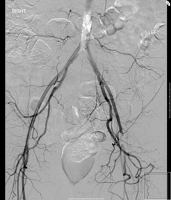 An angiogram is a special X-ray test of the blood vessels. It is done to find out if your blood vessels are diseased. The commonest disease is called peripheral vascular disease or atherosclerosis and as its name suggests mainly affects the arteries in the periphery of the circulation eg the legs.
What is peripheral vascular disease?
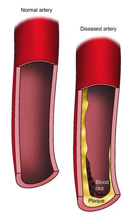
This is when cholesterol and fat builds up in your arteries. This build up of fat and cholesterol causes the artery to become narrowed or blocked. If blood can not flow through the artery it reduces the blood supply to that area and can cause you to have symptoms such as cramping, pain in your arms, legs or abdomen.
Why do I need an angiogram?
 Arteries supply blood to the organs and muscles in your body. An angiogram allows the doctor to see if there is a blockage or narrowing in a blood vessel that may interfere with the normal flow of blood though the body. These arteries can become partly blocked or completely blocked from a build up of cholesterol, cells or other material. This therefore reduces the blood flow to part of your body supply by those arteries e.g. your legs. An angiogram will provide your doctor with information to see if you need treatment such as angioplasty, bypass surgery or whether you can be treated with tablets. Your doctor may recommend an angiogram to diagnose a variety of problems with blood vessels including:
- Blockages of the arteries outside of your heart, called peripheral vascular disease
- Enlargement of the arteries, called aneurysms
- Abnormally formed arteries, called vascular malformations
- Kidney artery conditions.
Where will the procedure be done?
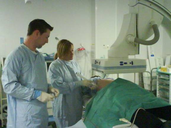
In a special room in the X-ray (radiology) department which is designated for this type of procedure. These vary in size and function from a small room to a large multipurpose operating theatre like that below. They are variously called interventional theatres, angio suites or cath labs.
How do I prepare for an angiogram?
You will usually be given a bed in the hospital. You may be asked to arrive in the hospital the day before the procedure or on the day of the procedure.You will be asked not to eat approximately 6 hours before the procedure. You will be given a hospital gown to wear. You will be seen by a doctor, the test will be explained to you and you will be asked to sign a consent form. This is to ensure you understand the test and its implications. The procedure is generally carried out using access via an artery in the groin. You may therefore be asked to shave the skin around this area.Please let the doctor know if you have any allergies. Please let the doctor know if you have had any previous reactions to X-ray (the dye used for kidney x-rays and CT scans). If you are diabetic on diabetic pills, you may be asked to not take these diabetic pills on the day of the procedure. If you are on any regular medication, please bring this in to hospital with you so that the doctor has a list of your medication which you take. Please inform your doctor if you are pregnant. The doctor or nurse may put a needle into your arm so that you can have some sedation or painkillers during the procedure.
What happens during an angiogram?
 The exact technique described below may vary from patient to patient but the general outline is as follows:
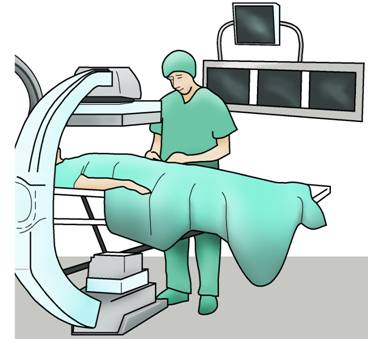
You will be positioned on your back, lying flat on the X- ray table. The team will ask your name and your date of birth. This is required for all medical procedures to keep our patients safe. You will have a monitoring device attached to your chest and finger, and may be given oxygen via a small tube in your nose or via a mask.The radiologists will keep everything as sterile as possible and may wear a theatre gown and theatre gloves. You will asked to keep your arms by your side. Usually the radiologist will decide to do the procedure using a large artery in your groin (but sometimes an artery near your wrist, elbow or knee may be used).
The skin around the groin will be cleaned with antiseptic (this can sometimes feel cold but will not be painful). The rest of your body will be covered by a large sterile sheet.The radiologist will place some local anaesthetic via a needle over the artery in your groin. The radiologist will then make a small nick in the skin. A needle is then inserted into the artery. A fine plastic tube called a catheter is then placed over the wire and into the artery. The radiologist will watch all of this on a tv-like monitor. A special clear X-ray dye called contrast, is then injected through the catheter. This dye allows the blood vessels to be visible on the X- ray and will show up any narrowed areas or blockages in the artery. The radiologist will then take the required X-rays. You may be asked to hold your breath for 5 -15 seconds while some of the X-rays are taken. If you wish, you can see the X-ray pictures on the screen during or after the test. When the radiologist is satisfied that the X-rays show all the information required, the catheter is removed. You might have a small amount of bruising in the groin. The radiologist will either press firmly on the skin entry – site for several minutes or place a small plug into the artery where the needle punctured the artery called an “angioseal”. This is done to prevent any bleeding from the artery.
Is it painful?
When the local anaesthetic is injected into the skin you may feel some discomfort. After this, the procedure should not be painful but you may have the sensation of gentle pushing and pulling in your groin. There will be a nurse standing nearby looking after you during the procedure. If the procedure becomes painful, please inform the nurse or doctor so that they will be able to give you some painkillers. As the dye is injected into the arteries you may feel hot (usually only lasts 20 seconds), a flushed sensation from the dye or the urge to urinate.
How long does the procedure take?
This varies from patient to patient. The length of time also depends on the location of the artery and how complex the procedure is. For example, if we are looking at a large artery in the leg, it can take 30 minutes, however if it is a small artery it may be more complex and can take longer i.e. one hour. As a guide, expect to be in the X-ray room for 2 hours.
What happens after the angiogram?
You will be taken to the ward on a trolley bed. The nurses on the ward will carry out regular observations such as pulse and blood pressure measurements, to ensure that there are no problems after the procedure. They will also look at the skin entry site in your groin. You will have to stay in bed for a few hours after the procedure. During this time, you should keep the leg or arm that was punctured straight to reduce bleeding from the puncture site. You may be allowed to leave home on the same day or be kept in overnight. You should not drive immediately after an angiogram. Therefore you should arrange for someone to take you home. You should resume normally activity after 2 days of the procedure.
What are the possible risks / complications?
An angiogram is generally a safe procedure, however there are some risks and potential complications:
(1) Bruise – This can occur around the site where the needle has been inserted into the artery. This is relatively common and is normal. You may also be tender in this region for a couple of days.
 (2) Haematoma – if there is bleeding from the areas where the catheter was placed, you may have a patch where blood collects under the skin called a haematoma. This usally clears up on its own.
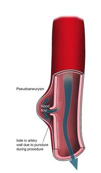
(3) False aneurysm – Rarely a pulsating lump develops in the groin at the site of the puncture. This is because bleeding occurred after the catheter was removed. The clot produced a small sac on the side of the artery via the hole made in the artery. This lump is connected to the artery and therefore has a pulse. The sac is called a false aneurysm. If the sac is above a certain size it is treated by a simple injection into the sac to block the small hole connecting it to the artery. This occurs in approximately 1 in 1000 people.
False aneurysm
(4) Infection- There is a risk of the puncture wound becoming infected. This can be treated with antibiotics
(5) Reactions to the dye – This is very uncommon. Various reactions and allergies can occur. The staff are fully trained and equipped to deal with such reactions. Reactions that can occur include rash, vomiting, asthma, disturbance in heart beats and kidney damage.
(6) Failure to do the procedure – As with any procedure, there is always a risk attempts may fail.
(7) Embolism or dissection – Occasionally damage to the artery occurs as a result of the passage of the instruments. This can result in bits of fatty material or blood clot being dislodged and blocking the artery further down the leg, or a splitting of the layers of the artery wall causing a blockage of the artery. These rare complications can normally be resolved by small procedures undertaken through the catheter but rarely a surgical operation will be required to restore blood flow.
When do I go home?
On the night of your procedure. It is important that you rest completely until the next day to ensure the puncture site heals up. The next day, you can have a shower but try and keep the skin site dry. If it gets wet, just pat it lightly down with a towel. You should avoid any heavy lifting for 48 – 72 hours after the procedure. You may notice a small bump under the skin, at the puncture site. This bump may last several weeks but will eventually disappear. However, if you have any concerns, you are advised to see your doctor. Some hospitals provide a day case angiography service to suitable patients and you may be able to go home within a few hours of the angiogram.
How will I get the results of the angiogram?
The scans will be looked at by the radiologist, who will write a report to the referring doctor. The report is usually available immediately in urgent cases or within 14 days of the procedure in elective (non-urgent) cases.